


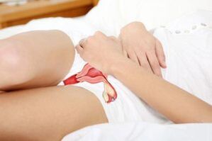
Varicose veins of the pelvis, unlike a similar disorder on the lower extremities, are not always noticeable, but are very harmful to health.Can lead to deterioration of blood flow in internal organs and disruption of their function.
Difference between varicose and healthy veins
The wall of a healthy vessel is always in good shape - this allows it to maintain blood flow.
The walls contract, although not as much as in the arteries.This effect is enhanced by contractions of smooth and skeletal muscles.
All veins located below the heart, i.e.in the limbs, abdomen, pelvis, chest, they have valves on their walls.They are “pockets” formed by the vascular endothelium.
They are located in such a way that blood flowing to the heart passes unhindered.With retrograde movement, it fills the valves and closes the vessel.
Blood always flows from the periphery to the heart, even against gravity.
Varicose veins undergo a number of changes in the valve apparatus.They reduce the tone of the wall and increase its permeability.This creates conditions for blood stagnation.
Some of the fluid bypasses, causing overflow of healthy vessels.And also provoking tone disturbances already in them.
What is it and why is it dangerous?
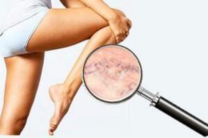
Varicose veins can affect peripheral vessels in any area of the body.
Manifestations of pathology on the legs are most noticeable - this creates a significant aesthetic defect.But the same phenomenon in the abdominal or pelvic organs is asymptomatic for a long time.

It is most dangerous for the female reproductive organs, less often for the bladder.Varicose veins of the rectum manifest as hemorrhoids.
Varicose veins of the pelvis can have various causes.The disease leads to disruption of blood flow in the reproductive system, and as a result, a deterioration in its function.
A woman may notice a change in the menstrual cycle, pain, and signs of hormonal disorders.Physical impact may result in vessel rupture and bleeding.
Reasons
Refers to multifactorial, i.e.arises due to a combination of a number of reasons.Among them:
- hereditary predisposition;
- lack of physical activity;
- pregnancy complications;
- overweight;
- diseases of the urinary system and intestines;
- hard physical labor.
The presence of one or even several unfavorable factors does not mean that the pelvic veins are dilated.This indicates a high risk of such a pathology.
Women with children are more susceptible to it than men.
The first signs usually appear around age 40 or in late pregnancy.The actual onset of pathology occurs much earlier.
Degrees
There are three degrees of development of varicose veins:
- Mild degree - damage to one or more peripheral vessels of the genital organs.Most often asymptomatic, may be temporary, requiring repeated examination.
- Medium degree - expansion of large veins of the parametrium - the outer lining of the uterus, or the myometrium - the muscular lining.Causes menstrual irregularities and poor health.
- Severe degree – varicose veins and swelling of most organs of the female reproductive system.Causes serious disorders in the sexual sphere, a high risk of disease and infertility.
Symptoms

Symptoms vary depending on the location and extent of the lesion.In the initial stages, it most often occurs without symptoms.
As the disease progresses, itching in the external genital area, heaviness and pain appear.
These sensations change in intensity depending on the phase of the menstrual cycle.They may intensify during bleeding and weaken a few days after it.
A woman may notice that her periods have become more painful and heavy.Sexual intercourse becomes painful, problems arise with conception and pregnancy.
Diagnostic methods
There are several ways to identify the disease.
The first and simplest of them is a gynecological examination.Identifies dilated veins on the skin, vaginal wall and cervix.Its data may indirectly indicate the cause - endometriosis, cervical erosion.
The most reliable method is gynecological ultrasound.Its advantage is that it allows the detection of dilation of the parametrium, fallopian tubes and ovaries.The results of this study provide more complete information about the state of the reproductive system.
In controversial cases, an MRI, a vaginal smear, a blood test for sex hormones, and a diagnostic curettage are prescribed.
Which specialist should I contact for help?
Treatment is carried out by a gynecologist in collaboration with a vascular surgeon.In case of hormonal disorders, consultation with an endocrinologist is necessary.
Since the disease is multifactorial, other specialists can also join in its treatment if necessary.
Therapy methods
Before starting treatment, you should undergo a complete examination of the reproductive system.Identify possible pathologies.As a rule, the disease occurs against the background of other disorders.

For greater efficiency, an integrated approach is used.Several methods of therapy are used at once, which makes it more successful.
Drug treatment
Includes taking drugs that increase the tone of the vascular wall - Hesperidin, Diosmin.
To reduce the permeability of vascular wall tissue, the following is prescribed:

- Ascorbic acid;
- Nicotinic acid;
- Rutin.
Their use is permissible even during pregnancy and after childbirth, if the first symptoms appeared then.
In addition, the doctor may recommend blood thinning medications - acetylsalicylic acid, vitamin K.
During pregnancy, their use is permissible only after consultation with an obstetrician-gynecologist, if the benefit outweighs the possible harm.
Compression and sclerosing therapy
Compression tights and belts are worn to prevent complications of varicose veins.They create pressure on the vessels of the external genitalia, preventing them from overflowing.
Due to this, normal or close to normal venous outflow is maintained throughout the pelvis.The degree of compression is determined by the doctor.You can wear such underwear during pregnancy.
Sclerotherapy is a procedure for introducing a special drug into the vessels that stimulates a short-term inflammatory response.And then - complete obstruction of the damaged vein and its transformation into a constriction.The blood flow in it completely stops.
This procedure can be done in the 2-3 trimester of pregnancy, if there are no contraindications.
Operation
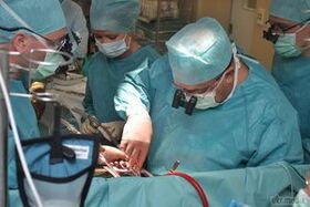
Surgical treatment is the removal of the affected area.It can be performed in several ways depending on the patient’s condition.
In some cases, it is necessary to perform partial resection of the ovary.And sometimes complete removal of the uterus of the fallopian tubes.
In women of childbearing age, doctors try to preserve the reproductive organs.
Therapeutic exercise
These are exercises that help improve blood flow and reduce venous stagnation.The most effective:
- "birch";
- back arching in the knee-elbow position;
- exercises for raising the legs and the sacral area.
They promote the outflow of blood due to gravity.
Folk remedies

Traditional methods of treatment are less effective than therapy in a hospital, however, they can significantly improve the patient’s well-being.
For this purpose, baths with medicinal plants are used - willow, oak branches, cudweed grass and chamomile.
They have a calming effect, reduce tension and stimulate blood flow.
Prevention recommendations
It is impossible to completely exclude all factors affecting venous outflow.But you can reduce their influence.For example, make time for exercise and maintain your weight within normal limits.
Prevention of the disease during pregnancy is of particular importance.During this period, be sure to monitor both nutrition and physical activity.And also for a sufficient amount of vitamins.